
How to Cite
Copyright notice

This work is licensed under a Creative Commons Attribution-NonCommercial 4.0 International License.
Copyright (c) 2026 Elifnur Sunger, Beyza Kalkanli, Veysi Yildiz, Tales Imbiriba, Giovanna Guidoboni, Peter Campbell, Deniz Erdogmus
Keywords
Abstract
Purpose: Accurate estimation of blood vessel tortuosity from medical images is an extremely important and challenging task. It is particularly relevant in the context of retinopathy of prematurity (ROP), where the staging of disease severity and consequent therapeutic approaches are heavily informed by the presence and prominence of vessel tortuosity. Existing methods based on centerline or skeleton curvature fail to capture curvature gradients across a rotating tubular structure, thereby limiting their effectiveness in the case of ROP.
Design: A methodological study with validation on artificial and real images.
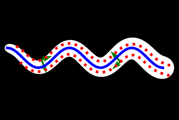
Methods: This paper defines local tubular curvature and presents the Tubular Curvature Filter (TCF) method, which locally calculates the acceleration of curve bundles traversing a tubular object parallel to its centerline. These curves are parallel to the centerline in the sense that they are integral curves following the direction of the ridge. This is achieved by examining the directional rate of change in the eigenvectors of the Hessian matrix of a tubular intensity function in space. TCF implicitly calculates the local tubular curvature without the need to explicitly segment or extracting the centerline of the tubular object.
Results: Experimental results demonstrate that TCF provides accurate estimates of local curvature at any point inside tubular structures. Results on two- and three-dimensional images show that TCF discerns curvature differences between the inner and outer sides of curved tubular objects, while centerline-based approaches cannot.
Conclusion: Our findings highlight that TCF’s ability to discern between the inner and outer sides of curved tubular objects is particularly useful in medical fields that require vasculature curvature analysis from images, especially where vascular structures often have non-uniform diameters, such as in ROP.
References
Deng H, Zhang W, Mortensen E, et al. Principal Curvature-Based Region Detector for Object Recognition. 2007 IEEE Conference on Computer Vision and Pattern Recognition. 2007; 1–8. https://doi.org/10.1109/CVPR.2007.382972
Thanh DN, Prasath VS, Hien NN, et al. Melanoma skin cancer detection method based on adaptive principal curvature, colour normalisation and feature extraction with the ABCD rule. J. Digit. Imaging. 2020;33(3): 574. https://doi.org/10.1007/s10278-019-00316-x
Mokhtarian F, Suomela R. Robust image corner detection through curvature scale space. IEEE Trans. Pattern Anal. Mach. Intell. 1998;20(12): 1376–1381. https://doi.org/10.1109/34.735812
Zhang W, Sun C, Breckon T, et al. Discrete curvature representations for noise robust image corner detection. IEEE Trans. Image Process. 2019;28(9): 4444–4459. https://doi.org/10.1109/TIP.2019.2910655.
He X, Yung NHC. Corner detector based on global and local curvature properties. Opt. Eng. 2008;47(5): 057008. https://doi.org/10.1117/1.2931681.
Tang Y, Li H, Sun X, et al. Principal curvature measures estimation and application to 3D face recognition. J. Math. Imaging Vis. Oct. 2017;59(2): 211–233. ISSN: 0924-9907. https://doi.org/10.1007/s10851-017-0728-2.
Fischer P, Brox T. Image descriptors based on curvature histograms. German Conference on Pattern Recognition. Sept. 2014; 239–249. ISBN: 978-3-319-11751-5. https://doi.org/10.1007/978-3-319-11752-2_19.
Qiuxiang Z, Yin K, Duan Y. Image reconstruction by minimizing curvatures on image surface. J. Math. Imaging Vis. Jan. 2021;63. https://doi.org/10.1007/s10851-020-00992-3.
Zhong Q, Li Y, Yang Y, et al. Minimizing discrete total curvature for image processing. Proceedings of the IEEE/CVF Conference on Computer Vision and Pattern Recognition. 2020; 9474–9482. https://doi.org/10.1109/CVPR42600.2020.00949.
Lee SH, Seo JK. Noise removal with Gauss curvature-driven diffusion. IEEE Trans. Image Process. 2005;14(7): 904–909. https://doi.org/10.1109/TIP.2005.849294.
Kim S. PDE-based image restoration: A hybrid model and color image denoising. IEEE Trans. Image Process. 2006;15(5): 1163–1170. https://doi.org/10.1109/TIP.2005.864184.
Nc SK, Radhika Y. Optimized maximum principal curvatures based segmentation of blood vessels from retinal images. Biomed. Res. (Tokyo), 2019;30, 308–318. https://doi.org/10.35841/biomedicalresearch.30-19-068.
Lutton JE, Collier S, Bretschneider T. A curvature-enhanced randomwalker segmentationmethod for detailed capture of 3D cell surface membranes. IEEE Trans. Med. Imaging, 2021;40(2): 514–526. https://doi.org/10.1109/TMI.2020.3031029.
Goldluecke B, Cremers D. Introducing total curvature for image processing. 2011 International Conference on Computer Vision. IEEE. 2011; 1267–1274. https://doi.org/10.1109/ICCV.2011.6126378.
Chen D, Mirebeau JM, Cohen LD. A new finsler minimal path model with curvature penalization for image segmentation and closed contour detection. Proceedings of the IEEE Conference on Computer Vision and Pattern Recognition. 2016; 355–363. https://doi.org/10.1109/CVPR.2016.45
El-Zehiry NY, Grady L. Contrast driven elastica for image segmentation. IEEE Trans. Image Process. 2016;25(6): 2508–2518. https://doi.org/10.1109/TIP.2016.2545244.
Gilbert C, Foster A. Childhood blindness in the context of VISION 2020: the right to sight. Bull. World Health Organ. 2001;79(3): 227–232. Available from: https://pubmed.ncbi.nlm.nih.gov/11285667/.
Chiang MF, Quinn GE, Fielder AR, et al. International classification of retinopathy of prematurity. Ophthalmology. 2021;128(10): e51–e68. https://doi.org/10.1016/j.ophtha.2021.05.031.
Goldman R. Curvature formulas for implicit curves and surfaces. Comput Aided Geom Des. 2005;22(7): Geometric Modelling and Differential Geometry, 632–658. ISSN: 0167-8396. https://doi.org/10.1016/j.cagd.2005.06.005.
Gong, Yuanhao and Tang, Wenming and Zhou, Lebin and Yu, Lantao and Qiu, Guoping. A discrete scheme for computing image’s weighted gaussian curvature. 2021 IEEE International Conference on Image Processing (ICIP). 2021; 1919–1923. https://doi.org/10.1109/ICIP42928.2021.9506611.
Taylor JE. II—mean curvature and weighted mean curvature. Acta Metall. Mater. 1992;40(7): 1475–1485. https://doi.org/10.1016/0956-7151(92)90091-R.
Choi G, Cheng C, Wilson N, et al. Methods for quantifying three-dimensional deformation of arteries due to pulsatile and nonpulsatile forces: Implications for the design of stents and stent grafts. Ann. Biomed. Eng. Dec. 2008;37, 14–33. https://doi.org/10.1007/s10439-008-9590-0.
Atiskova Y, Wildner J, Spitzer M, et al. Retinal vessel tortuosity as a prognostic marker for disease severity in Fabry disease. Orphanet J. Rare Dis. Dec. 2021;16. https://doi.org/10.1186/s13023-021-02080-0.
Bullitt E, Gerig G, Pizer SM, et al. Measuring tortuosity of the intracerebral vasculature from MRA images. IEEE Trans. Med. Imaging, 2003;22(9): 1163–1171. https://doi.org/10.1109/TMI.2003.816964.
Lorigo LM, Faugeras OD, Grimson WEL, et al. Curves: Curve evolution for vessel segmentation. Med. Image Anal. 2001;5(3): 195–206. https://doi.org/10.1016/S1361-8415(01)00040-8.
Law MW, Chung AC. Efficient implementation for spherical flux computation and its application to vascular segmentation. IEEE Trans. Image Process. 2009;18(3): 596–612. https://doi.org/10.1109/TIP.2008.2010073.
Han HC. Twisted blood vessels: Symptoms, etiology and biomechanical mechanisms. J. Vasc. Res. Mar. 2012;49, 185–97. https://doi.org/10.1159/000335123.
Hathout L, Do HM. Vascular tortuosity: A mathematical modeling perspective. J. Physiol. Sci. 2012;62, 133–145. https://doi.org/10.1007/s12576-011-0191-6.
Gerig G, Gouttard S, Corouge I. Analysis of brain whitematter via fiber tract modeling. The 26th Annual International Conference of the IEEE Engineering in Medicine and Biology Society. 2. IEEE. 2004; 4421–4424. doi: https://doi.org/{10.1109/IEMBS.2004.1404229}.
Ge S, Tian H, Zhang W, et al. Automatic bony structure segmentation and curvature estimation on ultrasound cervical spine images-a feasibility study. Journal of Physics: Conference Series. 2822. (1): IOP Publishing. 2024; 012023. https://doi.org/10.1088/1742-6596/2822/1/012023.
Zhou GQ, Zheng YP. Assessment of scoliosis using 3-D ultrasound volume projection imaging with automatic spine curvature detection. 2015 IEEE International Ultrasonics Symposium (IUS). IEEE. 2015; 1–4. https://doi.org/10.1109/ULTSYM.2015.0485.
Vogel P, Kampf T, Guggenberger K, et al. Robust centerline prediction for accurate vessel wall visualization of intracranial vessels in multi-contrast 3D MRI data. arXiv preprint arXiv:2209.01422, 2022. https://doi.org/10.48550/arXiv.2209.01422.
Aletti M, Gerbeau JF, Lombardi D. A simplified fluid–structure model for arterial flow. Application to retinal hemodynamics. Comput. Methods Appl. Mech. Eng. 2016;306, 77–94. ISSN: 0045-7825. https://doi.org/10.1016/j.cma.2016.03.044.
Koogler B, Young B, Campbell JP, et al. Analysis of vessel tortuosity and its impact on hemodynamics in retinopathy of prematurity. Investig. Ophthalmol. Vis. Sci. 2023;64(8): 1237–1237. Available from: https://iovs.arvojournals.org/article.aspx?articleid=2790163.
Javaheri B, Razi H, Gohin S, et al. Lasting organ-level bone mechanoadaptation is unrelated to local strain. Sci. Adv. 2020;6(10): eaax8301. https://doi.org/10.1126/sciadv.aax8301.
Ernst P, Hille G, Hansen C, et al. A CNN-based framework for statistical assessment of spinal shape and curvature in whole-body MRI images of large populations. Medical Image Computing and Computer Assisted Intervention–MICCAI 2019: 22nd International Conference, Shenzhen, China, October 13–17, 2019, Proceedings, Part IV 22. Springer. 2019; 3–11. https://doi.org/10.1007/978-3-030-32251-9_1.
Liu T, Yang Y, Wang Y, et al. Spinal curve assessment of idiopathic scoliosis with a small dataset via a multi-scale keypoint estimation approach. Adjunct proceedings of the 2020 ACM international joint conference on pervasive and ubiquitous computing and proceedings of the 2020 aCM international symposium on wearable computers. 2020; 665–670. https://doi.org/10.1145/3410530.3414317.
Wang H, Song Q, Yin R, et al. B-spine: Learning B-spline curve representation for robust and interpretable spinal curvature estimation. Proceedings of the AAAI Conference on Artificial Intelligence. 38. (6): 2024; 5381–5389. https://doi.org/10.1609/aaai.v38i6.28346.
Santamarina A, Weydahl E, Siegel JM, et al. Computational analysis of flow in a curved tube model of the coronary arteries: effects of time-varying curvature. Ann. Biomed. Eng. 1998;26, 944–954. https://doi.org/10.1114/1.113.
Ozertem U, Erdogmus D. Locally Defined Principal Curves and Surfaces. J. Mach. Learn. Res. 2011;12(34): 1249–1286. Available from: http://jmlr.org/papers/v12/ozertem11a.html.
Montiel S, Ros A. Curves and surfaces. 69. American Mathematical Soc., 2009; Available from: https://bookstore.ams.org/gsm-69-r/.
Ozertem U, Erdogmus D. Nonparametric snakes. IEEE Trans. Image Process. 2007;16(9): 2361–2368. https://doi.org/10.1109/TIP.2007.902335.
Law MW, Chung AC. Three dimensional curvilinear structure detection using optimally oriented flux. ECCV 2008: 10th European Conference on Computer Vision, Marseille, France, October 12-18, 2008. Proceedings, Part IV 10. Springer. 2008; 368–382. https://doi.org/10.1007/978-3-540-88693-8_27.
Law MW, Chung AC. An oriented flux symmetry based active contour model for three dimensional vessel segmentation. ECCV 2010: 11th European Conference on Computer Vision, Heraklion, Crete, Greece, September 5-11, 2010, Proceedings, Part III 11. Springer. 2010; 720–734. https://doi.org/10.1007/978-3-642-15558-1_52.
Law MW, Garvin GJ, Tummala S, et al. Gradient competition anisotropy for centerline extraction and segmentation of spinal cords. Information Processing in Medical Imaging: 23rd International Conference, IPMI 2013, Asilomar, CA, USA, June 28–July 3, 2013. Proceedings 23. Springer. 2013; 49–61. https://doi.org/10.1007/978-3-642-38868-2_5.
Brown JM, Campbell JP, Beers A, et al. Automated diagnosis of plus disease in retinopathy of prematurity using deep convolutional neural networks. JAMA Ophthalmol. 2018;136(7): 803–810. https://doi.org/10.1001/jamaophthalmol.2018.1934.
Cervantes-Sanchez F, Cruz-Aceves I, Hernandez-Aguirre A, et al. Automatic segmentation of coronary arteries in X-ray angiograms using multiscale analysis and artificial neural networks. Appl. Sci. 2019;9(24): ISSN: 2076-3417. https://doi.org/10.3390/app9245507.
Van der Walt S, Schönberger JL, Nunez-Iglesias J, et al. scikit-image: image processing in Python. PeerJ, 2014;2, e453. https://doi.org/10.7717/peerj.453.
Zeng A, Wu C, Xie W, et al. ImageCAS: A large-scale dataset and benchmark for coronary artery segmentation based on computed tomography angiography images. Comput. Med. Imaging Graph. 2023; 102287. ISSN: 0895-6111. https://doi.org/10.1016/j.compmedimag.2023.102287.
Dalvit Carvalho da Silva R, Soltanzadeh R, Figley CR. Automated Coronary Artery Tracking with a Voronoi-Based 3D Centerline Extraction Algorithm. J. Imaging, 2023;9(12): 268. https://doi.org/10.3390/jimaging9120268.
Jain AK. Fundamentals of digital image processing. Prentice-Hall, Inc., 1989; Available from: https://dl.acm.org/doi/10.5555/59921.