How to Cite
Copyright notice

This work is licensed under a Creative Commons Attribution-NonCommercial 4.0 International License.
Copyright (c) 2026 Gal Antman, Irit Bahar, Alon Tiosano, Alon Harris, Yamit Cohen-Tayar, Yair Zimmer, Amoy Fraser, Mehul Patel, Iftach Yassur, Itay Gabbay, Yehonatan Weinberger, Keren Wood, Orly Gal-Or
Keywords
Abstract
Purpose: To describe novel early changes in ocular physiology following short-duration exposure to microgravity.
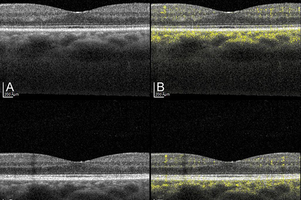
Observations: The subject was a 64-year-old astronaut who participated in the AX-1 mission that was sent to the International Space Station by AXIOM Space and the National Aeronautics and Space Administration (NASA) in April 2022 for 17 days. Comprehensive multimodal preflight and postflight ophthalmic examinations were performed, including anterior and posterior segment imaging and head magnetic resonance imaging (MRI). In addition, optical coherence tomography angiography (OCTA), which is not part of the standard NASA protocol, was conducted for the first time in this setting in the medical literature. Automated image processing was used to quantify flow signal pixels from the retina or choroid for separate analysis. The subject reported a new-onset need for reading glasses while in space. Mild widening of the optic nerve sheaths was found on MRI. OCTA studies demonstrated a significant postflight decrease in macular choroidal flow signals by 29% in the right eye and 11% in the left eye, and in retinal flow signals by 8.5% and 6.5%, respectively (p < 0.0001 for both factors for both eyes). Focal alterations were noted in choroidal thickness and Haller’s vessel diameter, which did not reach statistical significance.
Conclusion: A significant decrease in macular choroidal and retinal blood flow was observed in an astronaut after a short-duration spaceflight. These changes may serve as possible biomarkers of spaceflight-associated neuro-ocular syndrome and warrant further investigation. We recommend that future spaceflight evaluations include OCTA in the standard protocol.
References
Nelson ES, L. Mulugeta L, Myers JG. Microgravity-induced fluid shift and ophthalmic changes, Life (Basel). 2014;4:621-65. https://doi.org/10.3390/life4040621
Mader TH, Gibson CR, Pass AF, et al. Optic disc edema, globe flattening, choroidal folds, and hyperopic shifts observed in astronauts after long-duration space flight. Ophthalmology. 2011;118:2058-2069. https://doi.org/10.1016/j.ophtha.2011.06.021
Lee AG, Mader TH, Gibson CR, et al. Spaceflight associated neuro-ocular syndrome (SANS) and the neuro-ophthalmologic effects of microgravity: A review and an update. NPJ Microgravity. 6 2020;6:7. https://doi.org/10.1038/s41526-020-0097-9
Ong J, Tarver W, Brunstetter T, et al. Spaceflight associated neuro-ocular syndrome: Proposed pathogenesis, terrestrial analogues, and emerging countermeasures, Br J Ophthalmol. 2023;107:895-900. https://doi.org/10.1136/bjo-2022-322892
Stern C, Yücel YH, Zu Eulenburg P, Pavy-Le Traon A, Petersen LG. Eye-brain axis in microgravity and its implications for Spaceflight Associated Neuro-ocular Syndrome. NPJ Microgravity. 2023;20:56. https://doi.org/10.1038/s41526-023-00300-4
de Carlo TE, Romano A, Waheed NK, Duker JS. A review of optical coherence tomography angiography (OCTA), Int J Retina Vitreous. 2015;1:5. https://doi.org/10.1186/s40942-015-0005-8
Meiburger KM, Salvi M, Rotunno G, Drexler W, Liu M, Automatic segmentation and classification methods using optical coherence tomography angiography (OCTA): A review and handbook. Appl Sci. 2021;11:9732. https://doi.org/10.3390/app11209734
Corvi F, Corradetti G, Parrulli S, Pace L, Staurenghi G, Sadda SR. Comparison and repeatability of high resolution and high speed scans from spectralis optical coherence tomography angiography. Transl Vis Sci Technol. 2020;9:29. https://doi.org/10.1167/tvst.9.10.29
Lei J, Pei C, Wen C, Abdelfattah NS. Repeatability and reproducibility of quantification of superficial peri-papillary capillaries by four different optical coherence tomography angiography devices. Sci Rep. 2018;8::17866. https://doi.org/10.1038/s41598-018-36279-2
Schmidl D, Garhofer G, Schmetterer L. The complex interaction between ocular perfusion pressure and ocular blood flow - relevance for glaucoma. Exp Eye Res. 2011;93:141-155. https://doi.org/10.1016/j.exer.2010.09.002
Huang AS, Stenger MB, Macias BR. Gravitational Influence on Intraocular Pressure: Implications for Spaceflight and Disease. J Glaucoma. 2019 Aug;28(8):756-764. https://doi.org/10.1097/IJG.0000000000001293
Longo A, Geiser MH, Riva CE, Posture changes and subfoveal choroidal blood flow. Invest Ophthalmol Vis Sci. 2004;45:546-551. https://doi.org/10.1167/iovs.03-0757